
Diorama 1

About the Artwork
This artwork was commissioned as a response to an EPSRC funded research project that is developing peptides that can specifically attack and break down the membranes of cancer cells, and the artwork was developed alongside the research team.
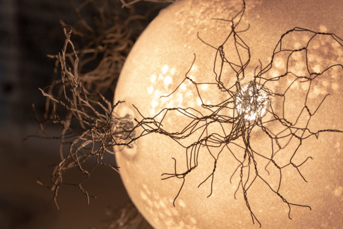
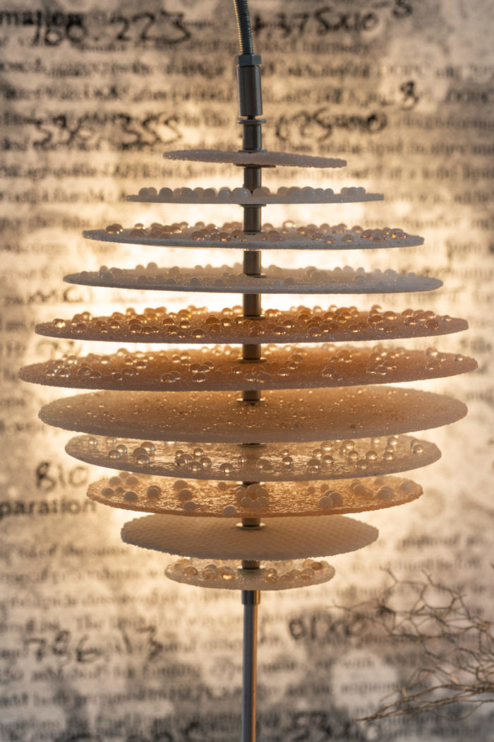
Diorama 1 references traditional dioramas, such as those found in natural history museums . It takes the form of an imagined microscopic landscape, populated by abstracted, cell-like structures set into a scientific research habitat that uses materials drawn from the research itself. Integrated lighting gradually changes through the day, creating variation and drawing attention to different features of the sculpture.
To find out more about the process and inspirations behind this artwork, read the artist's blog, Glass Bodies.
About the Artist
Julie Light is an artist working on themes of health and disease and visualising illness. She is interested in how people imagine their bodies internally and her work explores the implications of medical imaging, what it means to recognise your body from the inside out, and how that influences a sense of embodiment and identity. Julie holds an MA in Art & Science from University of the Arts, London, and her work has been exhibited across the UK in a variety of settings.
Alongside her individual practice, her collaborative projects have included The Museum of Extraordinary Objects and Changing Expectations of Art and Science with The Royal Society, and working with scientists at Imperial College and Kings College, London to create original artwork.
About the Science
In 2015, the Beales group and their collaborators published work investigating the interaction of the Polybia MP1 peptide with lipid membranes. This peptide is found in the venom of the Brazilian wasp Polybia paulista, where it has antibiotic properties. However this peptide had also been found to have selective anticancer properties. The new findings reported by Bueno Leite et al. in 2015 suggested a probable mechanism by which this peptide is more potent against malignant cells by targeting lipids that are known to redistribute to the outer surface of cancer cells and puncturing these membranes. This work received significant media attention, including the BBC, C&EN news and EurekaAlert!.
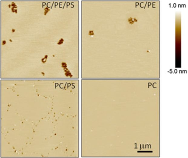
The MP1 peptide was found to have enhanced binding onto the surface of membranes containing PS lipids, but the lipid PE was required to facilitate the formation of large pores. Both these aminophospholipids have been reported to be present in enhanced concentrations on the surface of cancer cells, providing a synergistic mechanism for the MP1 peptide to preferentially rupture these membranes. The atomic force microscopy images above shows the pores (black) that form in membranes with different lipid compositions.
The current research aims to understand the relationship between the structure of this peptide and its ability to have selective interactions with a membrane dependent on its lipid composition. Using this physical insight, the team hopes to be able to design novel peptides with further improved potency and selectivity for cancer cell membranes that would make them amenable for development as a potential novel anticancer drug.
Read the 2015 Biophysical Journal research paper here.
Engaging with science through art
The rationale of commissioning an artwork about this research is to provide a gateway for the public to engage with the science on their own terms. Art allows the observer to develop a sense of intrigue that gives them the freedom to explore concepts with more freedom without needing to tackle specific scientific ideas or terminology that are unfamiliar to them. We hope this makes the science more accessible to a wider range of the general public, giving them a self-driven curiosity to frame their own ideas and questions and an inquisitiveness to want to find out more.
Funded by